Photoelectron Spectroscopy (PES)
1. The Experimental Contradiction of the Bohr Model
In 1913, Niels Bohr modeled electrons orbiting the atomic nucleus in fixed concentric shells ($n = 1, 2, 3\dots$). If the Bohr model had been complete, all electrons belonging to the same principal energy level (for instance, the 8 electrons in shell $n = 2$) would be located at the exact same average distance from the nucleus and have identical energy.
Under that hypothesis, ejecting any electron from the $n = 2$ shell would require the exact same quantity of energy. However, when multielectron atoms like Neon ($1s^2 2s^2 2p^6$) are ionized in a photoelectron spectrometer, electrons in the $n = 2$ shell produce two distinctly different energy peaks: one at $4.68\text{ MJ/mol}$ ($2s$) and one at $2.08\text{ MJ/mol}$ ($2p$).
2. Physics of the PES Spectrometer: Conservation of Energy
Photoelectron spectroscopy is the direct laboratory application of Albert Einstein's photoelectric effect to isolated, gas-phase atoms:
- Monochromatic Photons: High-energy photons of known frequency $\nu$ and energy $E_{\text{photon}} = h\nu$ (typically ultraviolet or X-rays) strike a gas-phase sample.
- Photoelectron Ejection: If $E_{\text{photon}} > \text{Binding Energy}$, a bound electron is ejected from the atom.
- Conservation of Energy Accounting:
E_photon = Binding Energy (BE) + Kinetic Energy (KE)
Binding Energy (BE) = E_photon - KE - Detector Measurement: The spectrometer measures the kinetic energy ($\text{KE}$) of escaping photoelectrons. Since $E_{\text{photon}}$ is precisely calibrated, the binding energy holding each electron to the nucleus is determined directly.


3. Reading the Spectrum: Jeremy Krug's Backwards Convention
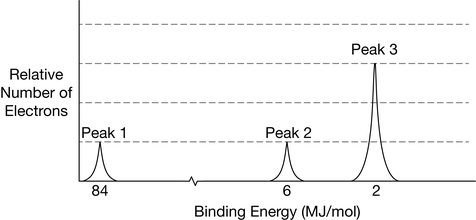
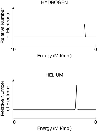
| Spectral Feature | Physical Meaning | How to Interpret on the AP Exam |
|---|---|---|
| Peak Position (x-axis) | Binding Energy ($\text{BE}$) | Identifies subshell identity ($1s > 2s > 2p > 3s > 3p$). Leftmost peak is ALWAYS $1s$. |
| Peak Height / Area (y-axis) | Relative number of electrons | Tells how many electrons occupy that subshell ($s = 2, p = 6, d = 10$). |
| Sum of Peak Heights | Total electron count | Equals atomic number ($Z$) for neutral atoms. |
4. Comparing Identical Subshells: Nuclear Charge Shifts
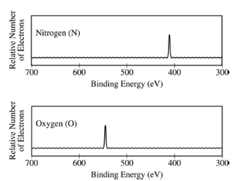
A classic AP Chemistry question compares identical subshells across different elements (e.g., the $1s$ peak of Nitrogen vs. Oxygen, or Silicon vs. Sulfur).
5. Step-by-Step Worked AP Exam Walkthrough Models
Problem Statement: A photoelectron spectrum for a neutral mystery element exhibits 4 peaks with the following characteristics:
- Peak 1: $104\text{ MJ/mol}$ (relative height = 2)
- Peak 2: $6.84\text{ MJ/mol}$ (relative height = 2)
- Peak 3: $3.67\text{ MJ/mol}$ (relative height = 6)
- Peak 4: $0.50\text{ MJ/mol}$ (relative height = 1)
Identify the element, write its complete ground-state electron configuration, and identify which peak corresponds to valence electrons.
- Identify subshells from left to right (highest to lowest energy):
- Peak 1 ($104\text{ MJ/mol}$, height 2) = $1s^2$
- Peak 2 ($6.84\text{ MJ/mol}$, height 2) = $2s^2$
- Peak 3 ($3.67\text{ MJ/mol}$, height 6) = $2p^6$
- Peak 4 ($0.50\text{ MJ/mol}$, height 1) = $3s^1$
- Total electron count: $2 + 2 + 6 + 1 = 11$ electrons.
- Element Identity: Neutral element with 11 protons/electrons is Sodium ($\text{Na}$).
- Valence Peak: Peak 4 ($3s^1$, $0.50\text{ MJ/mol}$) represents the outermost valence electron. It has the lowest binding energy because it is farthest from the nucleus and shielded by 10 core electrons.
Problem Statement: The $1s$ peak of Sulfur appears at $239\text{ MJ/mol}$, while the $1s$ peak of Silicon appears at $178\text{ MJ/mol}$. Justify this experimental observation using atomic structure and Coulomb's Law.
"Both Sulfur ($Z = 16$) and Silicon ($Z = 14$) have two electrons in their 1s subshell. Because these electrons reside in the n = 1 principal shell, the average distance (r) between the 1s electrons and the nucleus is approximately equal for both atoms. However, Sulfur possesses 16 protons in its nucleus, compared to only 14 protons in Silicon. According to Coulomb's Law ($F \propto \frac{q_1 q_2}{r^2}$), the greater nuclear charge in Sulfur exerts a stronger electrostatic attraction on the 1s electrons. Consequently, more energy is required to eject a 1s electron from Sulfur, producing a higher binding energy ($239\text{ MJ/mol}$ vs. $178\text{ MJ/mol}$)."